MICRO(NANO)FABRICATION AND MICRO(NANO)SENSORS
Micro and nanofabrication are flexible tools in permanent evolution that allow to explore the nanoscale realm of our world. At CNR-IOM we play with an exhaustive set of tools to design fabricate and test devices to detect photons, high energy electron beams, x-ray photons beams and THz radiation; we create microfluidics to interface wet chemical reactions and wet biology with such radiations; we develop nano-electro-mechanical systems and nanopatterned substrates mainly for biomedical applications, and more.
MECHANOBIOLOGY
Mechanical forces play many crucial roles in life, at different levels. At CNR-IOM, we are interested in the mechanical interaction at the cellular level:
Mechanotransduction, which is a process involved in various physiological conditions in mammals, such as conscious sensing of touch, pain, and sound, as well as unconscious sensing as proprioception. Mechanotransduction has also an important role in inflammation, tumor progression and metastasis.
Fertilization, differentiation, migration, embryology. From spermatozoon swimming into the egg, to the differentiation of the pluripotent cells into specialized cells. How these forces do their job is not fully understood, yet, and we’d like to found some answers.
The cell status itself is also characterized by its mechanical properties and represent a promising approach for medical applications.
Microscopy, optics, and biosensing. Visual information is the main channel for us, humans, to explore our world. However when going to micro and nanoscale naked eyes become useless. We develop new concepts in optical and electron microscopy to explore the living matter, and, why not, to develop sophisticated diagnostic tools.
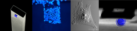
1) A high sensitive water compatible micromecanical sensor. 2) Hundreds of cell grown on a macrocantilever for parallelized
adhesion force measurement.3) A cancer cell imaged by AFM. 4) A mouse oocyte compressed by an AFM cantilever
CONTACTS
Dan Cojoc
CNR-IOM![]() cojoc@iom.cnr.it
cojoc@iom.cnr.it
Laura Andolfi
CNR-IOM ![]() andolfi@iom.cnr.it
andolfi@iom.cnr.it
Simone Dal Zilio
CNR-IOM
![]() dalzilio@iom.cnr.it
dalzilio@iom.cnr.it
Marco Lazzarino
CNR-IOM![]() lazzarino@iom.cnr.it
lazzarino@iom.cnr.it
Last updated on: 06/08/2020 - 12:31