The NanoInnovation Lab has expertise in: the physical-chemistry of organic thin films self-assembling; surface (bio)-functionalization and characterization; use of nanotechnologies for the controlled immobilization of confined, density-controlled monolayers of biomolecules for biosensing applications; single molecule interaction studies. Main topics of our research are:
- lipid bilayers of variable composition as mimicking platforms to study proteins/extra-cellular vesicles interactions with cell membranes
- development of high-sensitivity analytical platforms (Au-based nanoparticles; nanoarrays) for quantitative diagnostics and disease monitoring of molecular and vesicular biomarkers, as exosomes, in the context of cancer and neurodegenerative diseases
- tissue and cell biomechanics in health and disease
Towards these aims we use a combination of Atomic Force Microscopy (AFM) and AFM-lithography, fluorescence microscopy and synchrotron radiation based techniques. Also, we use advanced AFM imaging modes in liquid environment, to highlight structural details of protein/DNA interaction, at a single molecule level, and AFM force spectroscopy for studies in biomechanics.
The NanoInnovation Lab of Elettra is led by Dr. Loredana Casalis and Dr. Pietro Parisse. It is equipped with 4 state-of-the-art Atomic Force Microscopes (AFMs), one of which is mounted on a Nikon Eclipse Ti Inverted Microscope with a TIRF fluorescence setup and a CCD cooled camera, soon upgraded to single photon counting EM-CCD to habilitate super-resolution fluorescence imaging. We work in collaboration with other facilities/beamlines as The Structural Biology Lab, a fully equipped biological laboratory, including HT cloning, large-scale protein expression/purification equipment and different biophysical and biochemical approaches to characterize proteins and protein/protein interactions (FRET, UV-vis, etc.); vibrational spectroscopy beamlines (SISSI for Infrared Spectroscopy; IUVS for Raman Microscopy); microfabrication beamlines; the Small Angle X-ray Scattering beamline (SAXS), and of the free electron laser FERMI, as pump-probe experiments with ultra-short (fs) pulses.
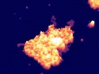
4µm x 3µm three dimensional rendering of Atomic Force Microscopy topographic map of extracellular vesicles
fused with a lipid bilayer mimicking a plasma cell membrane
CONTACTS
Loredana Casalis
Elettra-Sincrotone Trieste![]() loredana.casalis@elettra.eu
loredana.casalis@elettra.eu
Pietro Parisse
Elettra-Sincrotone Trieste![]() pietro.parisse@elettra.eu
pietro.parisse@elettra.eu
Last updated on: 06/08/2020 - 10:29